Figure 3 from Descriptive anatomy of the interscalene triangle and

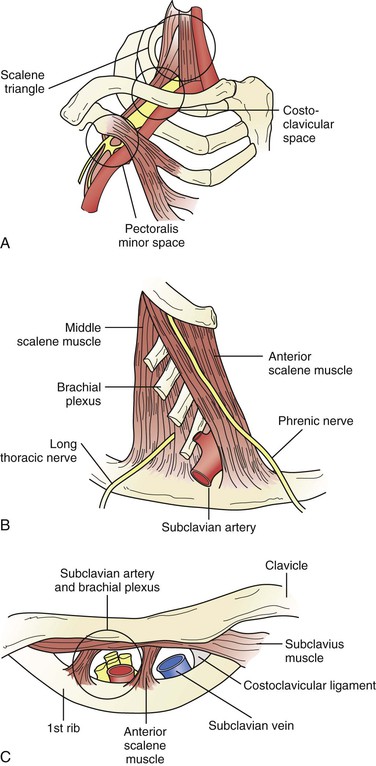
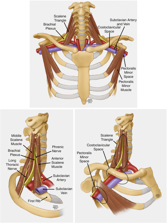
Fig 3. Depiction of the costoclavicular space. The neurovascular elements of the costoclavicular space can be seen here traveling superior to the first rib and inferior to the clavicle. The arrow indicates where measurements were taken. - "Descriptive anatomy of the interscalene triangle and the costoclavicular space and their relationship to thoracic outlet syndrome: a study of 60 cadavers."
RACGP - Neurogenic thoracic outlet syndrome

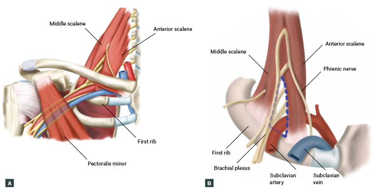
Thoracic Outlet Syndrome

Photograph showing the posterior triangle of the neck. The clavicular
Thoracic Outlet Syndrome

Daniel Clearfield, DO, MS, FAOASM on LinkedIn: Hydrodissection for the Treatment of Vascular Thoracic Outlet Syndrome

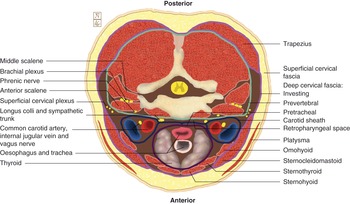
Posterior Cervical Triangle Lecture Flashcards
Surgical Techniques: Operative Decompression Using the Supraclavicular Approach for Neurogenic Thoracic Outlet Syndrome
Triangles of the neck: Anatomy, borders and contents
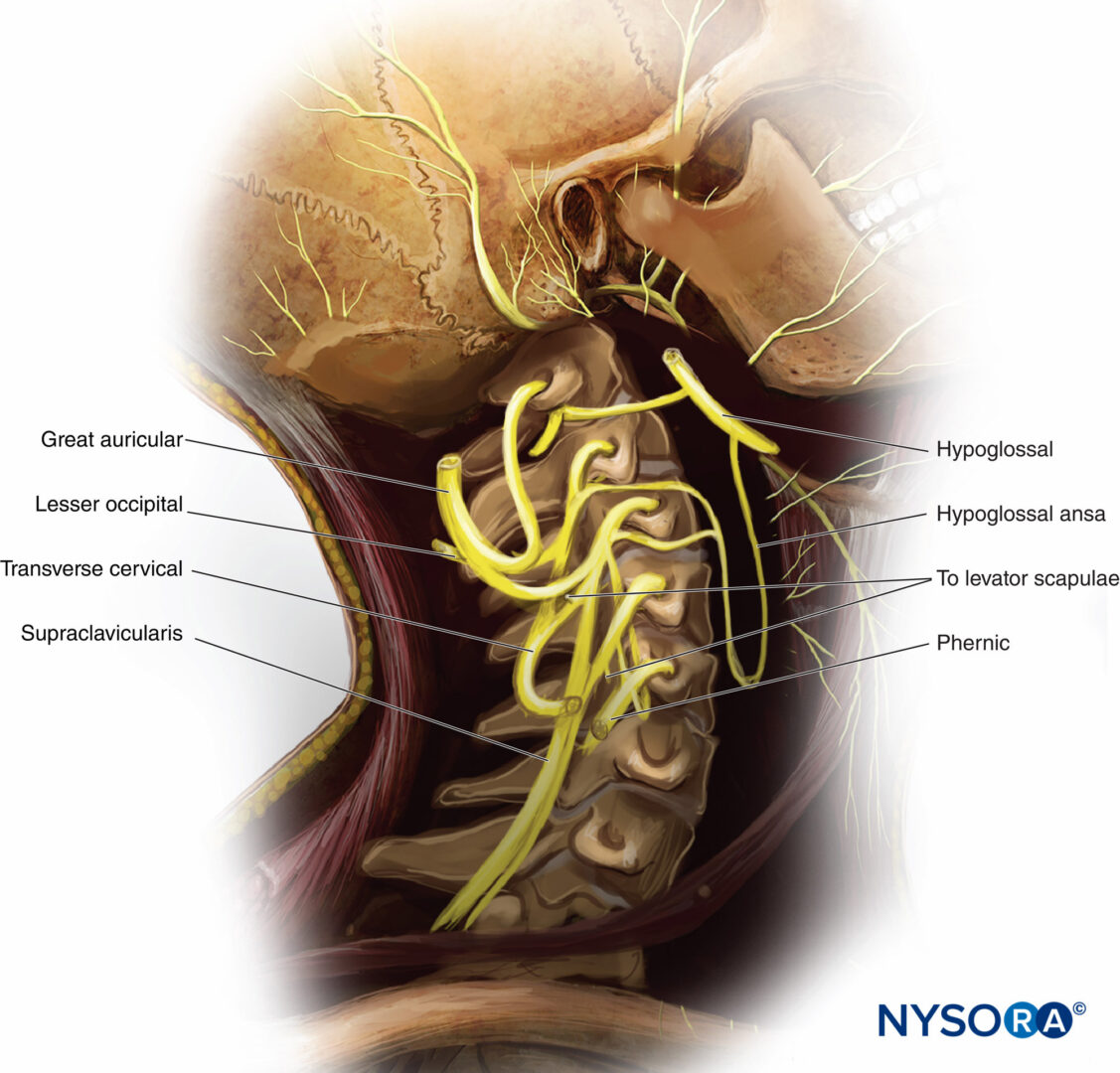
Functional Regional Anesthesia Anatomy - NYSORA
The neck (Chapter 4) - Applied Anatomy for Anaesthesia and Intensive Care

Schematic drawing of the triangles and anatomical structures in the

Medicina, Free Full-Text

Modern Treatment of Neurogenic Thoracic Outlet Syndrome: Pathoanatomy, Diagnosis, and Arthroscopic Surgical Technique - ScienceDirect
Figure 3 from Descriptive anatomy of the interscalene triangle and the costoclavicular space and their relationship to thoracic outlet syndrome: a study of 60 cadavers.