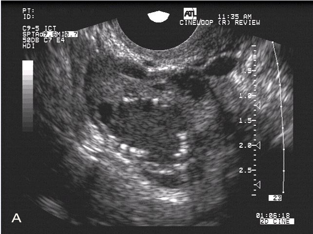
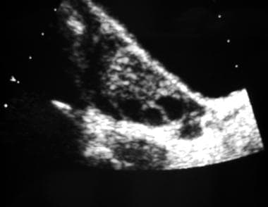
A–C) Sonograms of the dominant follicle (Ø 22, 32 and 38 mm) in a


Management factors affecting reproductive performance and causes of infertility of Ardi goats in Saudi Arabia - ScienceDirect

A–C) Sonograms of the dominant follicle (Ø 22, 32 and 38 mm) in a

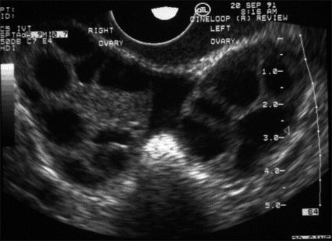
Sonogram of follicles in both ovaries. Left sonogram shows follicles
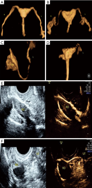
The value of transvaginal 4-dimensional hysterosalpingo-contrast sonography in predicting the necessity of assisted reproductive technology for women with tubal factor infertility - Gu - Quantitative Imaging in Medicine and Surgery

Follicle Monitoring and Endometrial Correlation

PDF) Artificial insemination in the anoestrous and the postpartum white rhinoceros using GnRH analogue to induce ovulation
Ultrasound in Follicle Monitoring for Ovulation Induction/IUI
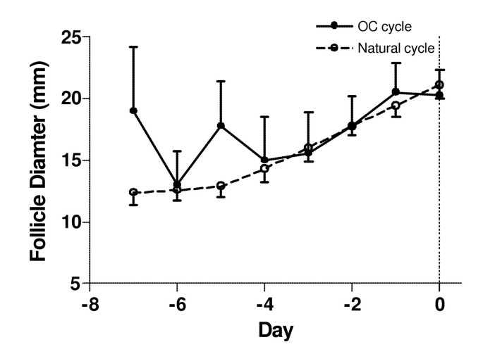
Ultrasound image attributes of human ovarian dominant follicles during natural and oral contraceptive cycles, Reproductive Biology and Endocrinology

Potential factors result in diminished ovarian reserve: a comprehensive review, Journal of Ovarian Research
Transvaginal ultrasonography and female infertility
Polycystic Ovarian Syndrome: Practice Essentials, Background, Etiology

Ultrasound Evaluation of Ovaries