Figure, B-Mode ultrasound showing main portal] - StatPearls
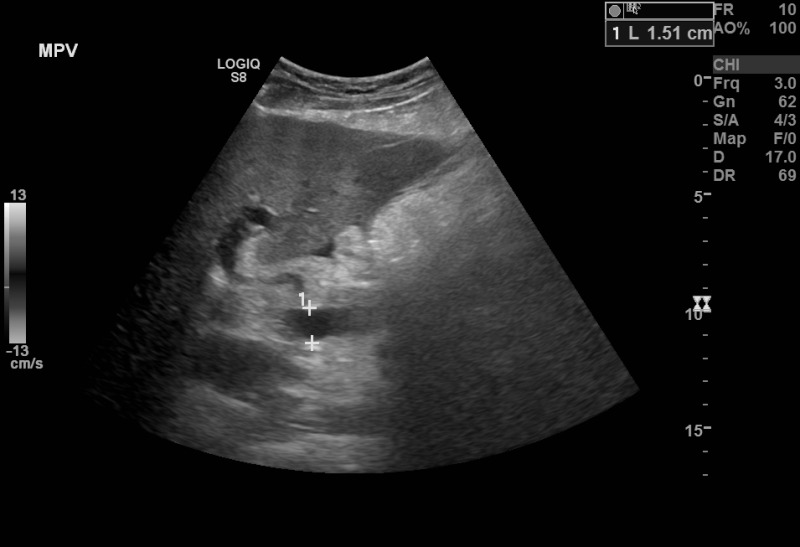
B-Mode ultrasound showing main portal vein diameter of 15.1 millimeters. This is an indirect finding of portal hypertension. Contributed by Brian Covello, MD
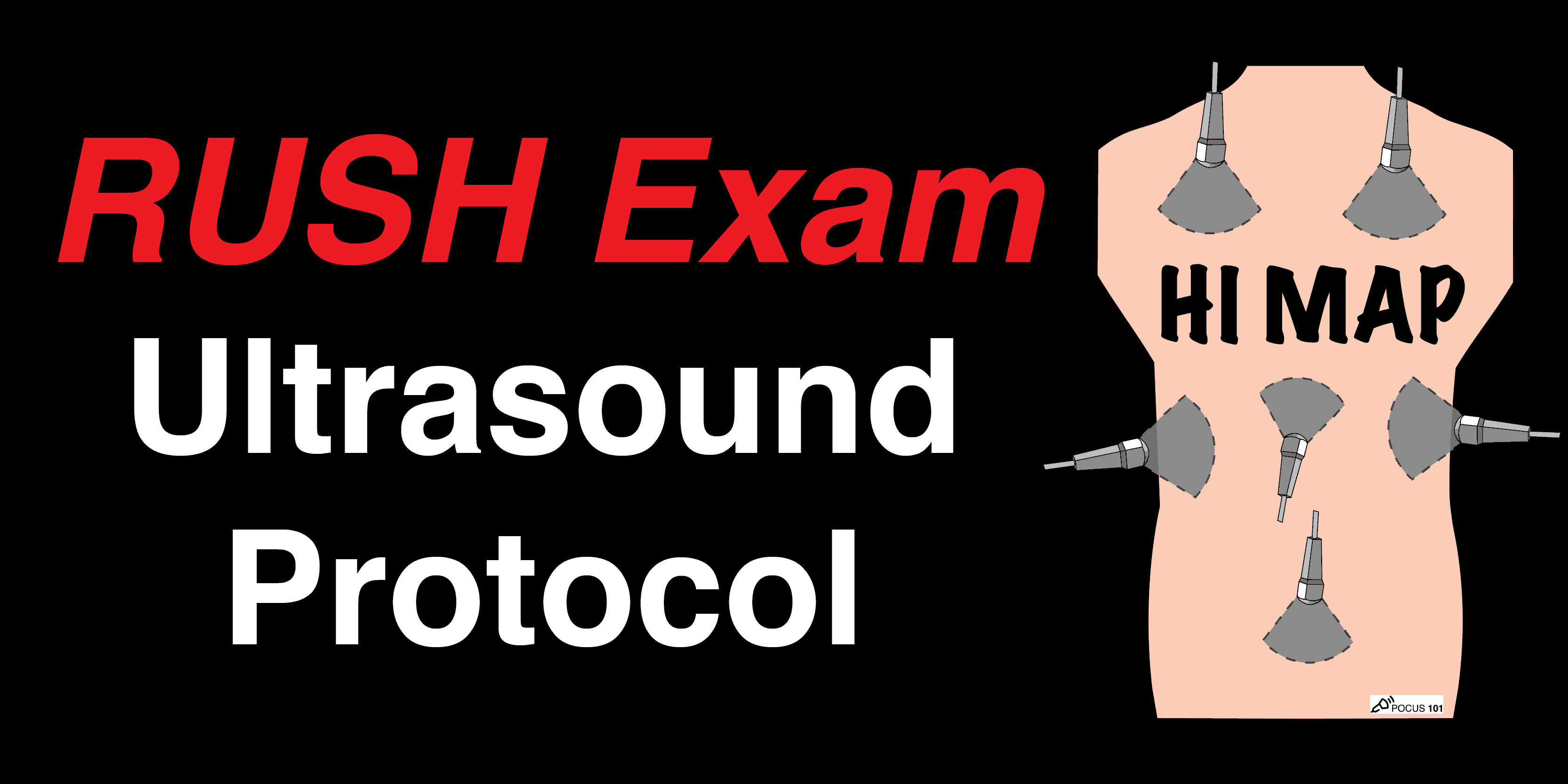
RUSH Exam Ultrasound Protocol: Step-By-Step Guide - POCUS 101

PDF) Ultrasound characteristics of abdominal vascular compression syndromes
Giant cell tumor with additional synovial proliferations [April 2023] – EFSUMB

PDF) Sonography 2nd Trimester Assessment, Protocols, And Interpretation

Ross Hauser, MD Reviews Cervical Spine Instability and Potential Effects on Brain Physiology
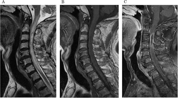
Intramedullary spinal cord abscess involving Actinomyces and Streptococcus: a case report and literature review

Diagnostics, Free Full-Text

Assessment of the portal vein anatomy with 3 D ultrasound
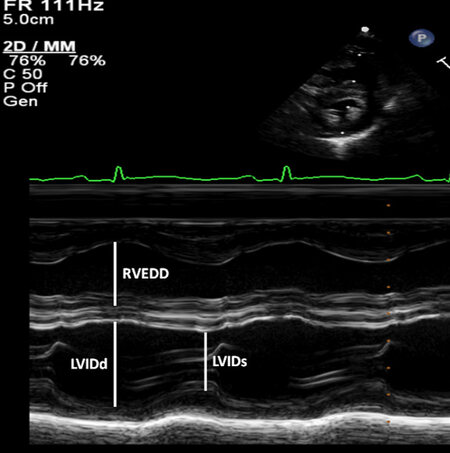
Echocardiography: an overview - part I

Gallbladder sludge, Radiology Case

Ultrasound B-mode examination of the 4-chamber (A, B) and 3-vessel (C

– Emergency Medicine EducationSplenic Infarction: ED Presentation, Evaluation, and Management - - Emergency Medicine Education
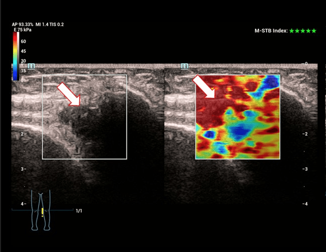
B mode ultrasound image (A) shows the hypoechoic tract (arrow) which is